
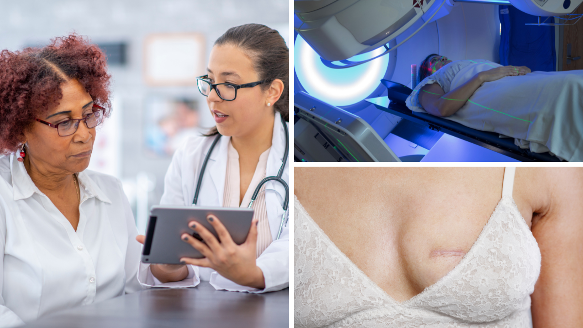
If you or someone you care about has recently been diagnosed with early-stage breast cancer, you likely have a lot of questions—and we’re here to help you find answers. Join us for a free, live webinar to learn more about two of the most common treatments: breast surgery and radiation therapy.
On this page
Breast surgeons and radiation oncologists often work together to create a treatment plan personalized to your needs. During this session, you’ll hear from experts Nicole Simone, MD, and Kristin Brill, MD, FACS, as they walk through the roles of lumpectomy, mastectomy, and radiation—and how they decide which treatments to recommend and when to begin them.
We’ll also cover the latest advancements in breast cancer surgery and radiation oncology, and most importantly, attendees had the opportunity to ask their questions live.
Whether you’re navigating your own early-stage diagnosis or supporting someone who is, this is your chance to get trusted information—directly from the people shaping the future of breast cancer care.
Watch or listen to the recording, or download the presentation slides below.
About our speakers
Nicole Simone, MD
Professor & Vice Chair, Research and Faculty Development of Radiation Oncology, Associate Director, Cancer Research Training and Education, Sidney Kimmel Cancer Center; Radiation Lead, Jefferson Breast Care Center
Dr. Simone is a physician-scientist at Thomas Jefferson University. Her NCI-funded laboratory explores how caloric restriction, or a reduction in overall calories, can make chemotherapy and radiation more effective, slowing tumor growth and spread while improving survival in breast, prostate, and lung cancer models. She brings her research directly to the clinic and currently has two open clinical trials using dietary strategies for people with breast cancer and brain metastases.
Read more
Kristin Brill, MD, FACS
Associate Professor, Enterprise Surgeon Leader for Breast Surgery; Director, Breast Surgery Program, Sidney Kimmel Cancer Center
Kristin L. Brill, MD, FACS is board-certified surgical oncologist with comprehensive range of services for patients with malignant and benign disease, including breast-conserving surgery, nipple-sparing mastectomy, preventive mastectomy, reconstruction, accelerated partial breast radiation device placement, and clinical trials for breast cancer. Dr. Brill has a particular focus on young women with breast cancer and people at high risk for breast cancer.
Read moreAbout our moderator
Caroline Koffke, RN, BSN, OCN
Director, Educational Programs, LBBC
Caroline Koffke, RN, BSN, OCN, is an experienced oncology nurse with a focus on breast cancer care. Over the years, she has honed a compassionate, patient-centered approach, providing expert guidance and support to individuals navigating the complexities of breast cancer treatment. As the director of educational programs, Caroline leads initiatives to develop and deliver high-quality, evidence-based educational resources for patients, caregivers, and healthcare professionals.
Read moreStay connected
Sign up to receive emotional support, medical insight, personal stories, and more, delivered to your inbox weekly.
Transcript
Caroline Koffke, RN, BSN, OCN (00:00:08):
We are so fortunate tonight because we have two amazing physicians joining us. I’m honored to introduce both of them to you, Dr. Nicole Simone, and Dr. Kristin Brill.
(00:00:18):
Dr. Simone is a professor and vice chair, research and faculty development of radiation oncology, associate director of cancer research training and education at Sidney Kimmel Cancer Center. She’s also the radiation lead at Jefferson Breast Care Center.
(00:00:33):
Dr. Brill is an associate professor, enterprise surgeon leader for breast surgery, and the director of the breast surgery program at the Sidney Kimmel Cancer Center.
(00:00:43):
As you can tell from their bios, they’re both absolute phenoms in their fields, and they work very closely together at Jefferson. Both are based here in Philadelphia. Thank you to both Dr. Brill and Dr. Simone for joining us. And for more information, you can read their full bios on our website.
Kristin Brill, MD, FACS (00:01:01):
I’m very excited to talk about cancer surgery. It’s changed so much. I’ve been doing this for about 24 years, and it’s so exciting to see some of the changes that are going on. So this was a nice opportunity. This is just a passion, you know, some of the changes and developments that we’ve, you know, identified and now that we can share with patients.
(00:01:24):
So, without further ado, I just want to do a little bit of background information just so we have an idea.
(00:01:30):
We’re talking about early breast cancer, and so just a reminder, baseline mammograms at 35 to 40. There are technologies that we have now to help, and there’s a lot of information now about dense breast and management of dense breast tissue. We’re still trying to get the word out and let women know that they are candidates for adjunct imaging like ultrasound, but there are even other, particularly for women who’s got increased risk, other modalities that we can use to help sort through dense breast tissue.
(00:02:01):
So this is a passion for our team and I think for all of us. One of the technologies I like a lot — in addition to tomosynthesis, that 3D mammogram — is contrast enhanced mammogram. It’s sort of like MRI, but it’s a mammogram that is performed with IV contrast. So it sort of gives functional imaging, meaning that it looks for areas that light up in the breast, in addition to anatomic findings, like masses and calcifications. In addition to that, of course, there’s breast MRI. We use that in high-risk screening and newly-diagnosed women.
(00:02:42):
Mammogram is not foolproof. We can miss up to 10% to 15% of malignancies on mammogram. That tends to happen more often in those women with dense breast tissue. So the sensitivity is a little bit less for a younger woman who may still have dense breast tissue. As we get older, our tissue becomes less dense with lack of hormonal influence.
(00:03:06):
Breast cancer is the most common cancer among women. It is very survivable. We’re here to talk about early breast cancer, but you can see the number of women diagnosed per year: 310,000. That’s up just a bit, about 1%. We’re seeing about 1% per year, but it does fluctuate. And overall, it stays pretty steady. Lifetime risk is about 13%, or 1 in 8 is the number that we’re all familiar with. Interesting to remember that about 1% of all breast cancer is diagnosed occur in men, just under 3,000 per year.
(00:03:42):
The average age is still around 61 or so; that’s the most common age for diagnosis. And this is one of my favorite slides. It always has been. I still share this with patients. It really does help when you get a pathology report or you’re trying to understand why there’s discussion about atypia on a biopsy, what DCIS is, what invasive cancer is, and that continuum from atypia that can go on to DCIS and invasive cancer.
(00:04:11):
I’ll say that most atypical cells, those lining cells of the ducts, which is where almost all breast cancers begin. Most atypia does not go on to progress into breast cancer, but we do recognize it as a tangible risk marker and one that we can actually modify with risk-reduction hormone therapy. So that’s something we frequently talk to women about when we see increased risk, especially with atypia.
(00:04:38):
I love this slide always because I think when women come in, especially with a favorable and early-stage breast cancer, and we talk about, “Well, it may have been there for 5 or 6 years,” they’re shocked. They feel like they haven’t done a good job getting their mammogram, maybe it’s not been done well. But breast cancer is extremely slow growing. So from the inception, when we’re talking about at a cellular level, until it gets to the size that it can be detected first on imaging, it takes several years. So that actually, I see that as opportunity to identify breast cancer. And really that’s where our screening comes in. Again, you know, standard screening and then adjunct or more sensitive screening for those women at high risk.
(00:05:21):
So breast cancer diagnosis, the most common cancer is invasive ductal. We also see invasive lobular, and DCIS is that early-stage, stage 0, or we used to call precancer years ago. We recognize it as a cancer, but it’s treated a little bit differently and it picked up a little bit differently.
(00:05:43):
I think Caroline hit on one point that I think is really important to remember. This is a big team of people. When a woman comes in, starting off with her mammogram, diagnosed with a cancer, goes on to meet a breast surgeon, maybe a plastic surgeon, a medical oncologist, the radiation oncologist, and then many other really important members of the team. And I saw a lot of them on our call tonight, genetic counselor, behavioral med, our nurse navigators are phenomenal, nutritionists, and physical therapists. It is a village, and take advantage of every piece of that because there is benefit to every piece of this as you go through.
(00:06:25):
So just just a review of stage, what early-stage breast cancer is. And that’s, you know, typically stage 0, stage I, and up to stage II and III. Stage IV is metastatic disease. But early stage is typically 0 through II, and that’s when the tumors are relatively small. Might be multifocal, but smaller tumors with limited lymph node involvement.
(00:06:51):
When a woman is diagnosed, there are a couple of key things that help dictate the course of treatment even for early-stage breast cancer. One of the important things, as many of you know, is hormone receptor status. Is the tumor, if we’re looking particularly at an invasive cancer, is it estrogen and progesterone sensitive? We like to see that tumors are highly estrogen and progesterone sensitive, as those normal ductile cells from which those tumor cells arise are very highly estrogen and progesterone sensitive. So the higher that number, the more that tumor has retained normal functions and characteristics. So that’s actually a good thing. Sometimes women think that they’re producing too much estrogen or there’s some problem, but that’s actually a favorable feature.
(00:07:39):
We look to see if the tumor is HER2 sensitive or HER2 positive. That puts us down typically a different path, sometimes with chemotherapy first, even with an earlier breast cancer, with a targeted therapy and some protocols that do put us on a different path. We recognize over a certain size, we will bring in HER2-based chemotherapy for that patient either before or after surgery.
(00:08:11):
Triple-negative is a particular variant that it’s not hormone receptor sensitive. It’s not HER2 positive. And so, again, over a a lower threshold size, triple-negative breast cancer is also treated with systemic chemotherapy either before or after surgery.
(00:08:30):
One of my favorite topics, of course, I’m a breast surgeon, so that makes sense: What we do to treat early-stage breast cancer.
(00:08:37):
Two basic options, right? Lumpectomy — and with lumpectomy is lymph node testing and some discussion on treatment with radiation, Dr. Simone is going to delve into that a little bit more. The other option with either a noninvasive DCIS or invasive cancer is mastectomy, and that can come in different varieties. A simple mastectomy, meaning just a straight mastectomy with no reconstruction. Skin sparing is actually saving the surrounding skin for immediate reconstruction typically. And nipple sparing is also for immediate reconstruction.
(00:09:14):
With mastectomy, the goal is to remove all breast tissue. We say routinely and recognize that it’s almost impossible to remove every speck of breast tissue, but it’s really quite complete. And so it’s a dramatic risk reducer, say, in a woman who is mutation positive or at higher risk.
(00:09:33):
At the same time as mastectomy, we do axillary node testing or sentinel node biopsy, and we’ll talk a little bit more about that.
(00:09:42):
You probably have heard a little bit more about hidden scar. This is something that has sort of evolved. When I started off as a breast surgeon, oh gosh, in the early 2000s, one of the features that got me really interested is I saw a lot of general surgeons doing breast surgery and taking out big wedges of tissue, and there was certainly less concern and attention to cosmetic result. Again, breast cancer is very survivable. We want, all women, but especially young women coming in for treatment to have their desired results. So even with lumpectomy, we try and hide the scar.
(00:10:24):
So you can see if we use that option around the nipple, safer, that tumor in the upper outer quadrant, we can actually tunnel and lift skin and get out there. Sometimes we go from an axillary incision that might be a little better hidden. Sometimes if the tumor is lower, we can use that IMF that lower incision and kind of lift the breast and get the tumor from underneath. So we can be pretty sneaky and get these tumors out through hidden incisions.
(00:10:50):
We try to make the incision as small and cosmetically placed as possible. We sometimes rotate tissue around to fill in a defect. But as Dr. Simone will mention, they love it when we put clips in behind where the tumor was so they know where to target radiation.
(00:11:11):
And then at the same time we map out for a sentinel node. And we’ll talk a little bit more about that, because sentinel node protocols have changed pretty dramatically in the last 10 to 15 years. So I want to talk a little bit more about that.
(00:11:31):
Every woman who’s diagnosed with a cancer has an option of lumpectomy and mastectomy. I should say, every woman who’s diagnosed with a breast cancer has the option of doing a mastectomy, may have the option of doing a lumpectomy. So there are some women who come in with a diagnosis, and we talk about management with lumpectomy versus mastectomy. They may be candidate for lumpectomy, but may ultimately choose mastectomy. But I wanted to just kind of run through how we guide patients on choice of surgery.
(00:12:03):
If a woman comes in with more extensive disease, especially if there’s an invasive cancer with a wider area of DCIS around it spanning more than 3, 4, 5 centimeters, depending on the size of the breast, that may be a better scenario for mastectomy than lumpectomy.
(00:12:21):
Sometimes if a woman has a very large breast and it’s more confined disease, it’s smaller and less DCIS, sometimes we’ll offer a reduction at the time of lumpectomy. And that’s a really nice procedure. Some women who are very large breasted who have always intended to do reduction, like that option. It’s covered by insurance and we can do reduction before they actually go on to radiation. If a woman has multifocal disease depending on her age, the extent the type of cancer she may very well be better treated with mastectomy than lumpectomy. Certainly women who are diagnosed with a gene mutation, and we know that their risk of having subsequent breast cancers is 40% to 60%, then mastectomy is discussed. And we often discuss bilateral mastectomy in that scenario.
(00:13:16):
If a woman’s had prior surgery and radiation, particularly if there’s recurrence and the lumpectomy bed are in the same region, we would think about mastectomy at that point.
(00:13:28):
I sometimes have discussions with patients who struggle with imaging. Extremely dense breast tissue, lots of findings — we do MRI, lots of things light up. There are some women who will opt to forgo close surveillance that would come with lumpectomy and opt to go with mastectomy.
(00:13:50):
Some women are not a candidate for radiation, and radiation is an important part of the treatment if we offer breast conservation, for example, scleroderma. And then probably one of the most common reasons is patient’s choice. They say: I just, I want to avoid radiation, or I want to avoid future MRIs or mammograms. I want to be complete. I don’t want to think about it. I tend to be a worrier. I am more comfortable going with mastectomy.”
(00:14:20):
So it does take a lot of kind of looking through options and some of the features that may guide a decision, and it takes a lot of discussion.
(00:14:28):
What I tell patients typically is, when they come in, it’s an information gathering session, and it’s not really intended for her to walk out and give me a definitive decision. So it does take some thinking. And I do encourage women not to make decisions immediately, because what you may decide, you know, 48 hours after a cancer diagnosis may not be what you really want to do long term.
(00:14:51):
So talking about types of mastectomies. Depending on whether we’re doing a skin sparing or nipple sparing mastectomy, or a simple mastectomy with no reconstruction. And that’s where no skin is saved and that the chest is flat down, typically with an incision across. But we work very closely with our plastic surgeons. So if we’re talking about doing a mastectomy there are different reasons we might offer nipple sparing versus skin sparing.
(00:15:18):
We try to offer nipple sparing, but two barriers to nipple sparing. One is the location and extent of the cancer. If we see cancer right behind that nipple-areolar complex, those ducks that all converge up at the nipple, and particularly if there’s a lot of DCIS, she may not be a candidate for nipple-sparing procedure. And MRI can help with that, mammogram can help with that.
(00:15:39):
If a woman has a larger and very ptotic, which is a really nice gentle way of saying droopy, breast and really the nipple is kind of down, kind of pointing down to the toes, that’s challenging to do nipple sparing. Typically, that’s a big cavity to be filled, and so that can be challenging. Although I want to show you a technique that we’ve been doing more and more often and having great success with for a woman who’s got some ptosis and a larger breast where we can actually reduce that skin envelope. You can imagine if we’ve got a woman with a very large breast that, you know, kind of hangs down onto the abdomen, to remove the breast tissue from underneath and fill that space in with either implant or tissue from the belly ends up being a really big reconstruction. It sits differently. It feels differently. It’s larger than her native breast.
(00:16:32):
This is that nipple-sparing, skin-reducing mastectomy, and it’s a little bit complicated. The idea is you see in that first panel, A, that gray area, the problem is blood supply to the nipple. We need to preserve blood supply to the nipple. So you see in A and B, we actually, the plastic surgeon will take the covering off the skin, what we call deepithelialize. So the outer skin layer, that keratinous layer on the outer, is kind of bladed off, but underneath, all these tiny little vessels are still preserving the nipple.
(00:17:15):
Eventually, once I’ve removed the breast tissue from underneath, and typically — you can see in panel C they’re putting an implant underneath — they maintain that blood supply to the nipple, and then they wrap that skin around. So it’s almost like a reduction incision, but preserving the blood supply to the nipple.
(00:17:33):
The possibility for nipple loss or inadequate blood supply is a little bit higher. We don’t see it often with a straight nipple-sparing mastectomy, but with this one, it can be a little bit more tenuous. It’s difficult to do when women who are smokers or have any of vascular disease. But this is a really beautiful procedure. And that final picture, it really does look that good. We just saw a patient in the office yesterday who had this done bilaterally, both sides, with an implant reconstruction. And honestly, you couldn’t tell that she had bilateral mastectomies. It’s just such a rewarding thing for patients.
(00:18:10):
One of the other really exciting evolutions over the last 8 years or so is the placement of the implant. When we do implant-based reconstruction, the tradition has been to do what’s on that third panel all the way to the right, subpectoral breast reconstruction. And so after the breast tissue was removed, the plastic surgeon will come on in and put an implant underneath the pectoralis muscle, and it was sort of held in place with that pectoralis muscle.
(00:18:45):
The downside to that is it required an expander upfront. You need a really more thick walled implant that is a temporary one that’s put in that has the ability to be expanded in the office over several weeks. So that expansion process, a little bit of saline injected in there week after week, can be really uncomfortable, especially, again, in young women who have more resilient pectoralis muscles. It’s uncomfortable, and you get a little bit of a blunted effect because the strong pectoralis muscle may be kind of blunting the implant a little bit.
(00:19:26):
What has been developed is this pre-pectoral breast reconstruction. And this has really brought some great options for us. So once the breast tissue was removed, which you can see in that middle panel that’s what’s happened, the implant is placed above the muscle, under the skin. The little bit of fatty tissue you see just under the skin is the normal subcutaneous fat that we need for skin viability and for the skin to do well. But the breast tissue has been removed. They use something called acellular dermal matrix. That’s that kind of light blue shaded layer. It’s a sheath of collagen. It’s actually a sheath of cadaver collagen. It’s iodinated, or ionized I should say, so that it is acellular meaning there are no cells, it’s just collagen.
(00:20:19):
So that is put into place, and that gets absorbed by that subcutaneous fat and all around, and eventually that gets absorbed all together and forms a new natural sheath around that implant to keep it in place. That has allowed us to go, what we call, direct to implant, meaning that in this situation, if it’s of the right size breast and the right situation, we don’t need to put a tissue expander in. We go directly to the permanent implant. Everything is done in one step. We do quite a bit of this in patients who are candidates. And that, in conjunction with the nipple sparing, is a really beautiful procedure. The incision is just underneath at the natural crease under the breast, and so there’s no visible incisions with this kind of reconstruction. And doing it in one phase without the expansion and above the muscle, much, much more comfortable.
(00:21:15):
The other sort of type, category of reconstruction that we offer is autologous flap. That means using a woman’s own fatty tissue to fill in that pocket that’s created after a mastectomy. In autologous flap, the most common one is coming from the abdomen. It’s sort of similar to a TRAM that we used to do years ago, but this is muscle sparing. TRAM was actually taking that rectus muscle, which you see down through the incision, that abdominal muscle.
(00:21:42):
We do not take the abdominal muscle now. We actually just did one of these today. This is one where the plastic surgeon, the microvascular surgeon, will find just the ideal pair of artery and vein that is perforating, supplying that fat pad, and they dissect that out. They leave a length on it so that it’s brought up under the breast flap that’s created once the breast surgeon has removed the breast tissue. And this also can be a nipple sparing. This diagram is not indicating nipple sparing, but it can be. And it’s connected into one of those branches that’s just between the ribs kind of under the breastbone that you see in that picture.
(00:22:26):
It’s a beautiful procedure. It is one and done. It’s a long procedure. It can be 10 hours or so, depending if we’re doing one or two sides. But it is nice because it offers it tissue under skin. And sometimes women say, who have been radiated in the past and have a lot of radiation change, this may be more ideal on a patient because when we put a woman’s own fatty tissue under skin, it’s a little bit more nourishing, the skin does better rather than foreign body like an implant.
(00:22:55):
So there are, you know, indications where this is a great reconstruction. It can be done one or both sides, depending on how much tissue that woman has on the belly. I think most of us know that we’ve been saving saving stuff on the belly for years for whatever we might need it for.
(00:23:11):
A little bit about breast anatomy, because I want to talk about sentinel node biopsy. The lymph nodes, 75% of the drainage goes to the axilla, about 25% of the breast drainage goes through the lymph nodes under the breastbone. But the lymph node staging becomes really important in terms of staging a cancer and helping to determine treatment. It goes into that whole algorithm on what form of radiation, what extent if a woman needs radiation at all, in whom we offer adjuvant chemotherapy, chemotherapy after surgery.
(00:23:54):
So this has changed quite a bit, and we’ve made some really great strides in terms of what we do for sentinel node analysis. It’s a little bit of a blurry slide. This is one of my older ones.
(00:24:12):
We generally inject either radioactive dye, that technetium 99 sulfur colloid, it’s a weak radioactive substance, back behind the nipple. Sometimes we use a blue dye, sometimes we use both. It’s injected not typically around the tumor but now back behind the nipple. And it takes about 30 minutes. That substance is taken up by the lymphatics behind the nipple and travels in the first one or two lymph nodes sort of in that whole chain. Typically, it’s a node a little bit lower down. There are about 15 or 20 lymph nodes in general. And those nodes are really defined anatomically. Those nodal chains extend on into the neck and into the chest. But we’re really interested to know if any tumor cells have gone to lymph nodes within the axilla. And so that’s the purpose of of testing. So it’s changed so much in the last 30 years.
(00:25:03):
It used to be, and not so long ago, certainly in the ’90s and up to about 2001 is when we abandoned, taking all nodes out with any invasive cancer. But if a woman in 1998 came in with a small early-stage, favorable invasive cancer, she typically got a complete axillary dissection. And there were some scenarios where the more nodes that were taken out, we thought, the better surgeon, you know, the better she may do. And it led to unacceptable rates of lymphedema, range of motion issues, numbness, chronic pain. Really some unsavory, you know, complications.
(00:25:48):
Sentinel node biopsy came around in the late ’90s, and by about 2001 most surgeons had done away with taking all nodes out but then mapping out that key one or two lymph nodes in the chain and testing those.
(00:26:03):
We used to, if there was a positive node, take them all out. Now there is even more data that came around about 10 years ago called Z0011. We all know it as Z0011, but it’s ACOSOG, which is American College of Surgeons Oncology Group Z0011 trial. This was a really pivotal study. It’s not one that even completed entirely. It was a meant to enroll about 1,900 patients. It closed early at 891. But in essence, this study told us that if a woman has negative lymph nodes before she goes to the operating room, and we identify a little bit of cancer in four or fewer nodes, that we don’t need to go on to take more lymph nodes out. That woman will be treated with something systemically; she’ll be treated with radiation. And taking all nodes out. And adding radiation and that systemic treatment, but particularly adding radiation, brings on more complications than it does benefit.
(00:27:10):
This has changed what we do entirely in terms of early-stage breast cancer and sentinel node biopsy, so this is a really pivotal study.
(00:27:25):
For early-stage breast cancer, NCCN guidelines, of course most of you know, this is like our bible. We go by this. And it’s fun to look and see as things evolve and change, it’s very current, how they recognize some evolutions in some of the treatments and it’s reflected in the guidelines. But in essence, when we have patients who have that hormone receptor-positive cancer after surgery, and we determine how risky that tumor is for systemic metastasis, she’ll be offered some anti-estrogen medication like tamoxifen, typically in premenopausal women, and aromatase inhibitors like Arimidex (anastrozole), and that’s in postmenopausal women.
(00:28:09):
For women who are node negative, and actually now in some in node-positive scenarios in postmenopausal women, we think about Oncotype. Oncotype I describe as a crystal ball, the best that we have, and we’ll talk a little bit more about that because that is a really amazing technology that we have to help determine adjuvant treatment after surgery.
(00:28:37):
For somebody who’s node positive, depending again on the menopausal status and the situation, particularly Oncotype diagnosis, we might consider chemotherapy. We also consider radiation, even in women who have had mastectomy.
(00:28:53):
So Oncotype is testing done on the tumor itself. And depending on whether a woman is premenopausal or postmenopausal, we might wait for surgical results. A woman who’s premenopausal and has a positive lymph node, she typically would be offered chemotherapy, and we would not send off Oncotype for testing. For a woman who is post-menopausal, if she’s got fewer than four lymph nodes positive, then we do think about sending off Oncotype.
(00:29:20):
It is a 21-gene expression panel. It looks at what we talked about already — ER, PR, HER2, Ki-67 — but it looks at another 15 genes and proteins. And this generates a score that helps us recognize who would benefit from systemic chemotherapy.
(00:29:45):
So Oncotype assumes — this is done on a woman Jane Doe. She is node negative. Her Recurrence Score is 13. She was born in 1951. She’s presumably postmenopausal. So if she takes 5 years of an anti-estrogen medication like tamoxifen or an aromatase inhibitor, her risk for recurrence at 9 years is 4%. The benefit she would get from chemotherapy is less than 1%. This woman would not be offered chemotherapy. There’s no benefit and only risk. All of her benefit is going to come from that aromatase inhibitor. So this is one of the tests we love to talk about and prepare patients for. It’s pretty amazing.
(00:30:33):
From a distant view, some of the things that have changed in the world of surgery and breast cancer treatment: Definitely improved techniques for detection. MRI, especially for dense tissue, and contrast-enhanced mammogram are great adjuncts. They have downsides, but they can be helpful in the right scenario. We are doing less surgery in the axilla and preserving lymph nodes. In women who are 70 and above who have very early, favorable cancers. We consider forgoing a lymph node biopsy altogether. We have emerging tools for characterizing tumors and risk of recurrence like Oncotype. And there’s some others very similar to Oncotype.
(00:31:14):
There is an expanded role for targeted therapies, and there is a whole other discussion on immunotherapy and targeted therapies that can be used before or after in the right scenario. And we’re continually defining the role of radiation. And I look forward to Dr. Simone’s talk about radiation. We work very closely, and of course our world’s overlap quite a bit.
(00:31:43):
And I think most exciting from the breast surgeon and plastic surgeon is a really dramatically improved cosmetic result and experience for patients.
Caroline Koffke, RN, BSN, OCN (00:31:53):
Dr. Brill, thank you so much. I feel like I just learned so much in in this short 25 minutes that you were explaining things to us. But I think the most impactful thing for me is just how much has changed in the right direction in surgery and how the advancements have led to better outcomes for individuals with this diagnosis. And you did such a nice job of highlighting that.
(00:32:16):
We have a ton of wonderful questions, some of which I actually think will be great for you and Dr. Simone to answer together. So I’m not going to ask all 11 that we have right now. I’m just going to pick one or two for you to answer before we go over to Dr. Simone.
(00:32:31):
But we did have a question from someone about the difference in survival for mastectomy versus lumpectomy. And I think this is a challenging one and kind of varies case by case, but I would love to hear your opinion on if there is or isn’t a difference in survival.
Kristin Brill, MD, FACS (00:32:47):
It’s a really good question and probably one that I thought about getting into, but it’s a really good question and a point that we should make.
(00:32:56):
For the most part, there is no difference in survival between lumpectomy and mastectomy. There can be a difference in local recurrence. So if we’re looking at it in early-stage breast cancer for a woman who, let’s say, has a favorable breast cancer, undergoing lumpectomy, some form of radiation, and goes on an aromatase inhibitor, her chance of local recurrence really should be on the order of 5% to 7%. For a woman undergoing mastectomy with or without reconstruction, her chance of local recurrence should be, not 0 but as close to 0 as we get, 1% or 2%. So there is a slight difference in local recurrence. In terms of systemic recurrence that’s really based more on lymph node status, tumor histology. So there’s no difference in treatment outside of, particularly, the possibility of a role of radiation. There’s no difference in treatment.
(00:33:51):
For instance, some women will say, “I’ve got this cancer, if I do a mastectomy, I’m less likely to need to have chemotherapy.” No. Because the rule for chemotherapy is really based on lymph node status, Oncotype, tumor type. That’s not changed by what you do at the level of the breast surgically.
(00:34:09):
So it’s true, there’s no difference in survival between the two procedures. And it’s a great point.
Caroline Koffke, RN, BSN, OCN (00:34:14):
Thank you so much. That’s actually unbelievable.
(00:34:16):
Another great question, and I’ll try to keep it brief, but what is the estimated post-surgery recovery time differences between lumpectomy, simple mastectomy, mastectomy with implant, DIEP flap?
Kristin Brill, MD, FACS (00:34:31):
Yep, it does vary. And so it’s interesting. Lumpectomy and sentinel node biopsy we typically do as a same-day procedure in the surgery center, even under just sedation, not even with general anesthesia. They go home the same day, there’s no drain, they’re a little sore and bruised, and the recovery is pretty simple. Generally we say about 2 weeks. Some women take a little bit more if they need it, if they have a very active job like motherhood.
(00:34:56):
For mastectomy and reconstruction, it’s longer. It can be a 6-week recovery, for that DIEP flap it can be 8 weeks. Especially for that DIEP flap, but also for implant-based reconstruction, no heavy lifting, no pushing, pulling for 6 weeks. So it really can, you know, limit activity, so that’s a little bit longer.
(00:35:19):
If it’s a two-stage reconstruction, meaning that tissue expander is put in at the beginning and they get exchanged to a permanent implant, in those scenarios where women are not a candidate for direct-to-permanent implant, that second stage, often women will go back to work for a couple of months and time out that second whenever it’s convenient for them. So usually about 3 months out.
Caroline Koffke, RN, BSN, OCN (00:35:41):
OK. Very interesting.
(00:35:41):
I definitely want to get to Dr. Simone, so then I can continue to pick both of your brains since I know you work closely together and can give a lot of advice to our constituents tonight. So thank you so much. We will bring you back on in a little bit.
(00:35:54):
But Dr. Simone, I would love to welcome you as our radiation oncologist and our expert in all things radiation to share your side of things.
Nicole Simone, MD (00:36:03):
Awesome, thank you. Thank you so much for having me, and thank everyone for attending when you could be probably doing some more fun things tonight.
(00:36:12):
I am going to talk a little bit about radiation. There’s a lot of things to talk about with radiation, and I’m going to try to be brief because I know everybody wants to get to questions as well. The one thing I really wanted to highlight tonight, and I will start getting into the meat and potatoes, is really talking about the differences in fractionation. I think that’s one of the more confusing things these days for patients, is that there’s a wide variety of things that are offered to patients at this time, and it can be very confusing and different centers offer different things. So I wanted to go through some of the data on what we call fractionation or the number of treatments that are offered.
(00:36:52):
I’ll talk a little bit about that and then talk about some innovative approaches that Dr. Brill and I are doing together at Jefferson, and things that, you know, people are doing around the country as well that are a little bit more innovative. I thought that might be of interest.
(00:37:05):
I do want to just talk about radiation in general. One of the most common things is for people to ask, What the heck is radiation? Is it going to burn me? What is it?
(00:37:14):
Radiation is something that is a global term, but when we talk about radiation therapy for patients, what we’re really talking about are x-rays. Just like you would get an x-ray like a chest x-ray or a mammogram. Those are very light x-rays. So they’re in what we call the kilovoltage range. They pass through the body, they stop at different things a little bit differently. So soft tissue, they’ll go a little faster; bone, it will change the direction. And so when you get an image that’s actually just reflective of how fast those x-rays were allowed to pass through the body.
(00:37:50):
When we treat with radiation to actually try to treat something in therapeutic radiation, those are actually just megavoltage radiation. They’re just heavier particles. And what that means is we can actually choose the dose and figure out where is something in the body — where’s the tumor in the body or the cavity in the body that we need to treat — and figure out what type of energy we should be using to deposit energy there. So they are still x-rays, meaning you don’t see anything, feel anything, or hear anything when you get something, nothing’s touching you, but those x-rays are able to deposit exactly where we want them to. And there’s a fancy planning system behind that. But that’s all it is. It’s just honestly x-rays.
(00:38:31):
They do cause DNA damage. So what happens is when a radiation beam is coming, sometimes it really kills in one of two different ways. It does damage cells, but the great thing about your normal tissues is those are actually able to repair themselves. They have DNA repair machinery. They just say, OK, all right, you know, just similar like to being, you know, in the sun. When you go outside, things get damaged and your body just repairs itself. When you have a cancer cell, the cancer cell does not have the same machinery. So when the cancer cell has exposure to radiation, it does one of two things. It either directly hits both of our DNA strands and causes direct kill, or it interacts with the water that’s right next to those cancer cells and hits that and it’s able to kill indirectly. So that’s how it mostly works. That’s kind of the global picture.
(00:39:32):
And so it is just x-rays, and that’s how it’s killing cancer cells preferentially to normal cells.
(00:39:40):
There are a number of different ways of delivering radiation. Most people talk about external beam radiation therapy. There has been some internal, you guys have probably heard of brachytherapy, maybe some of you have had that. Intraoperative radiation is another choice that we sometimes offer patients that we’ve had trials that Kris and I have worked on together with that. But most commonly people either have conventional x-ray radiation therapy or particle therapy. And some people have been offered protons, neutrons, or carbon ions even.
(00:40:12):
We just talked about a question someone had. I was going to get into that briefly, so I will pass right over that. As Kris was saying, the wonderful thing about data that we have is that these treatments are equally efficacious. So it really becomes a personal choice of a patient’s to decide what’s best for them after hearing everything that they have.
(00:40:35):
I am going to go through some data with you guys today, and the reason is I just feel like when you see the numbers in person right up in black and white, you can see that that things are really, the data that we have really shows equal efficacy.
(00:40:51):
This is really just showing breast conservation versus mastectomy. So this is exactly what Kris was saying. The rates of recurrence rates or death, those are exactly the same. We think that when you have just a lumpectomy versus a lumpectomy and radiation, that the radiation added to lumpectomy alone does provide a small amount of survival benefit.
(00:41:15):
One of the questions was talking about mastectomy versus lumpectomy with radiation. Those are equivalents, but sometimes we talk about just lumpectomy alone. Do we really need radiation? What’s it adding? My tumor was removed. What is radiation actually doing? We do think that without radiation there is a slightly higher chance of recurrence and that probably means that there’s microscopic cells that we can’t see, we can’t detect on imaging. And Kris went through that, how big something has to be when we are able to detect it on mammogram or MRI. So there’s probably a few cells hanging out, and that’s what can potentially cause the recurrence. That’s one of the reasons, and that’s why we think radiation works, is because there are potentially some cells around in the area that can be killed with radiation.
(00:42:04):
Can radiation be de-escalated? You’ve probably heard in the older times, if you have older relatives, patients had received between 5 and 6 weeks of radiation, standardly. That’s still something that’s done today for very young patients, some patients that have node-positive disease, some patients that we don’t have clear data on, but by and large, most patients are diagnosed initially with early-stage cancer. And so the question is do we really need 6 weeks of treatment or can we do something different.
(00:42:37):
So what are those principles? Well, we can talk about dose being reduced. Do we need as much radiation as we usually have? Or do we need to treat the whole breast? Can we just treat the part of the breast where the actual cancer was? We’ll kind of talk about some of those things. And then this really has to do with risk. Everything that Kris was talking about with choosing the proper surgery to really take care of cancers depending on hormone status, depending on lymph nodes, those all also play a role in the decisions of what type of radiation we should be getting. So tumor characteristics. How is the patient tolerating things? Do they have other diseases, like Kris was mentioning, maybe lupus or something else? How do we take into account all of those variables as well as the characteristics that are biological as well?
(00:43:27):
What is hypofractionation? People have probably heard that term, and that just means that you can kind of deliver a little bit less radiation total, but maybe a little bit more per day to achieve the same biologic equivalent. So really, even though we’re talking about fractionation or treatment numbers that are less than 30, they really are providing the same, what we call, biologically equivalent dose as the standard therapy that we had always used. And that’s how clinical trials are designed. That’s how fractionation is developed as we try to figure out exactly a number that will come up with the same biologic equivalent dose to get to what we want to.
(00:44:08):
Most people have now heard of 15 or 16 treatments for radiation for what we offer early-stage patients. And that’s based on two different trials. And these trials, when we look at early-stage breast cancer patients, Kris and I aren’t looking at what’s happening in 2 or 3 or 4 years. We really want to have some long-term data. We know patients with early-stage breast cancer will do well. We want to make sure that that whatever treatment we’re giving is a durable response. And so everything that we offer that is standard, it’s like, we can do this or we can do this. That’s always based on clinical trial data. And so we’re very thankful for people that have enrolled in clinical trials in the past. And it’s always based on equivalent outcomes or better outcomes at a longer time period, like 10 years, 15 years or 20 years.
(00:45:01):
And this actually is a great trial where they started with 50 gray in 25 treatments, that’s the 5-week regimen, and they compared it to 16 treatments, which is essentially 3 weeks and a day. But basically what I want to show you here is that when you look at recurrence and you look at survival, those things have exactly the same data. So whether or not we do the 3-weeks-and-a-day treatment for most patients, that’s going to be equal if you have early-stage disease. Now there’s a few things that your radiation oncologist may worry about. So if you have a lot of things that we may be concerned about, a high grade, something triple-negative, if there’s young age, maybe 40 years old. If you have a lot of those factors, you might not be a candidate, but it’s certainly something where I’m showing you that there is good long-term data.
(00:45:49):
I’m going to skip the start A trial and B trial. This is also over 2,000 patients. Again, we always have lots of patients on these trials. And they looked at, you know, 13 treatments or 15 treatments versus the standard. And again, we saw the same response rates. We also have noticed that most of them actually have excellent cosmesis. It’s almost virtually the same as in prior, the standard 5 to 6 weeks. And now the question is can we even go faster. Can we do five treatments? And there have been some wonderful clinical trials mostly out of Europe actually. So I’m going to just briefly talk about these because they actually have been in the news a lot in the last 2 years, and there is some great long-term data on this.
(00:46:35):
So this one, there’s about a thousand patients that are patients who are over 50, had a tumor less than 5 centimeters and randomized patients to the 5 weeks of treatment or once a week, just once a week, for 5 weeks. This is called the FAST regimen. And essentially what they found is equivalent results in both arms. So there was no change in local recurrence for either of those regimens.
(00:47:02):
Then there’s the FAST-Forward trial, and this is actually where you can deliver five treatments all in 1 week essentially, or 5 consecutive business days. So it was comparing what we now know, the 15 or 16 treatments that can be standard, with only five treatments. And they had two five-treatment regimens. One did better than the other one. So that’s what we standardly treat. And if your radiation oncologist is worried about either a close margin or a high-grade lesion, there’s the opportunity to give a little extra radiation for an additional five treatments. So this could be up to 10 treatments total. And what we found here too is that recurrence rates were exactly the same. The rates of distant relapse were exactly the same. So all cancer outcomes were exactly the same. And those things are definitely offered at centers and there’s great long-term data, at least 10 years on those trials.
(00:47:56):
This is another five-fraction regimen that’s now doing partial breast. So do we have to treat the whole breast, which is what I have been talking about up until now, or can we do partial breast? And I think Kris knows for a very long time, and I’ve been at treating breast cancer only for 17 years now. For a very long time I was not a fan of partial breasts because sometimes we saw some cosmetic outcomes that were not that great. If you’re only treating part of the breast with radiation but you have potential for getting some fibrosis or some scar tissue, it would be in one area. It was not my favorite.
(00:48:31):
But this actually, this trial has changed my opinion significantly. This was called the Florence trial that was done in Italy. What they did was they actually did external beam radiation, so they did not use brachytherapy. They delivered this in just five fractions, but actually every other day, not consecutive fractions, and did very small volumes and had not only excellent … and compared that to the standard 5 weeks. And they had not only excellent cancer outcomes at 10 years, but also excellent cosmesis. So this has actually become something that we offer a lot of patients if they’re a candidate for partial breast radiation.
(00:49:13):
And then the question is can you omit radiation altogether. I would love to say yes. I’m not a radiation lover. However, there really has not been a great population. We do have some data in older patients that endocrine therapy alone has a great chance of not having something recur after lumpectomy. But even in the trials with it are very specific: for patients over 70, tumors that are less than 2 centimeters, or estrogen-positive tumors. The radiation really has dropped local recurrence rates from about 10% at 10 years to 1%. So it’s still doing a great job locally. And a permanent local reduction could play an important role as we age.
(00:49:53):
So it would be nice, and we do have those balance discussions. Is this worth it to you? A 10% decrease? And we can talk through those things and actually kind of share in that decision-making process. Here’s the data, what do you guys want to do? And so that’s typically what we do.
(00:50:10):
I wanted to talk briefly about a collaboration that Kris and I have together. And I’m going to go through this really quickly. Talking about lifestyle change. Because people often ask when they’re diagnosed with cancer: What else can I do? How else can I help support myself?
(00:50:23):
It’s usually used in prevention. We think about lifestyle changes. We think about it in survivorship. But we actually think about it in terms of using food or diet as a drug. Can we actually help radiation work better? Our vision is really to try to empower patients to help get some additional response and feel good when they’re actually going through treatment.
(00:50:49):
What we’ve seen in models is that when you add caloric restriction or just a decrease in calories or a healthy diet to radiation, we can actually slow tumors down significantly. We also decrease the time to metastasis if that’s going to be something to occur. Again, these are model systems. And we actually improve the immune function. So we are able to increase our CD8 cells, which are really the T cell killer cells, and we can decrease the immune suppressive T cells.
(00:51:20):
So we actually have designed a clinical trial. We’ve done a few trials at Jefferson so far using diet with radiation, even just diet in a 2 to 3 week window before surgery alone. This current trial that we have open, it’s actually a randomized preoperative radiation trial. Now, preoperative radiation is something that’s being done around the country. And actually NRG one of the larger bodies that runs national cooperative group trials is designing a pre-op trial right now.
(00:51:51):
What we’re doing is we’re randomizing patients. We’re actually delivering — so you would have your diagnosis of breast cancer and we would actually talk together. You would meet with Dr. Brill, you would meet with myself, decide if you’re an ideal candidate. And what we can do is actually deliver radiation first before surgery, just five treatments to the partial breast. So it is the smaller area. Then half of our patients are being randomized to a dietary intervention. And our goal is really to see how how much does a tumor shrink? Does a tumor shrink?
(00:52:23):
So far we’ve enrolled 22 patients. It has shrunk in everyone so far. And what does the diet actually add to that? We are seeing some benefit with regard to increasing cell kill. So there’s actually fewer cancer cells when we’re adding diet to that radiation. So that’s our primary endpoint in our trial.
(00:52:44):
I’m going to skip some of these because we’re right at the end here. But these, you know, anyone who has a tumor less than 3 centimeters can enroll in the trial. If you’re over 40, you would also qualify. We do have a cutoff of looking at your BMI greater than 21 on this trial. And our goal really here — and I have these pretty pictures, but I don’t have time to go through them — is that when you deliver pre-op radiation, it’s actually a much smaller area that we deliver radiation to. And then you would go to surgery just a week or 2 after the radiation is over. So Dr. Brill essentially removes the tissue that has been radiated. And those are some pros, is that you’re not left with that radiated in your body anymore. And the only con that we have so far is that if you are found to have node-positive disease by Dr. Brill when she evaluates your sentinel node, if it wasn’t detected initially with ultrasounds, then you would need to have postoperative radiation.
(00:53:43):
So I’m going to skip ahead, but we do have great early data showing that we are getting some tumor shrinkage. People do feel better too. So we have had some quality-of-life studies showing that people feel a little bit better. And I’m going to just let you guys do some questions.
Caroline Koffke, RN, BSN, OCN (00:53:58):
We have so much important information, and I know there’s lots of questions for you as well.
(00:54:01):
I’m super interested in learning more about your clinical trial just because I think it is really amazing to see how we can amplify what you all are already doing with your science and technology with things that people can do at home as well. So that’s phenomenal. Thank you for sharing.
(00:54:18):
A couple questions specifically to you, and then I’ll invite Dr. Brill up again because there are some for the combination of you both.
(00:54:28):
Someone, I know this is also a common point. If anyone is node positive, let’s say it’s even just one node, are they getting radiation regardless of whether or not they’re getting a lumpectomy versus mastectomy?
Nicole Simone, MD (00:54:42):
So that’s dependent. Kris did talk a little bit about the Z0011 trial. And so sometimes with one node positive, we don’t need to, based on some of that data. It really depends on if there’s other adverse risk factors or if it’s a lymph node that’s still there after neoadjuvant chemotherapy for instance. There are a few risk factors that are nuanced, but no, one lymph node does not necessarily mean that you need radiation afterwards.
Caroline Koffke, RN, BSN, OCN (00:55:07):
Great. Thank you for clarifying that. Some other questions about, you know, obviously with radiation we’re damaging some DNA. Are there other organs that are impacted. What are some things that patients can do? Someone’s asking: Should I drink more water? Are there other things that patients can do to kind of prepare their body for this damage?
Nicole Simone, MD (00:55:24):
So that’s a great question. I think one of the things that we have learned significantly is that just a healthy, balanced lifestyle, if you can, during that time period. If you’re a smoker, please try not to smoke, because that can add to some of that. Even if you take a smoke break just for those few weeks, it’s actually been shown to help. Staying away from sugary foods has also been shown to help. And so those things may contribute. Just making sure that you’re getting rest as well so that your body does have the ability to repair that DNA damage.
(00:55:56):
And it’s hard, right? None of us sleep well to begin with. Just trying to set some good habits during that time to get your body in as good shape as you can.
(00:56:06):
And you asked a question about organs. So the nice thing about breast radiation is our breasts are on top of our body, right? So when we’re just treating the breast alone, we’re just kind of skimming across the surface of the chest wall. And so most of the time, especially using things like prone radiation or looking at breath hold in different ways that we can protect organs, we’re pretty anal about that. Just making sure that we are blocking out normal organs. Most of the time we actually do a really good job. And that’s also where partial breast radiation can come in to play, right? So if the tumor is in an ideal spot and we can do partial breast and be away from everything else, that’s a great place to be as well.
Caroline Koffke, RN, BSN, OCN (00:56:49):
That’s awesome. And I think it’s so worth noting that obviously this has come a very long way as well. Like I mentioned with Dr. Brill, in just the ability to have such targeted therapy, which is obviously what our goal is.
(00:57:02):
Another question, and again, you radiologists, you always get kind of, “Will this happen? Will this happen?” Someone’s asking: Can radiation cause skin cancer? Because this is a scary thing for people.
Nicole Simone, MD (00:57:15):
Yeah, of course. So anytime, we did talk about damaging DNA, anytime that repair process has to kick in, again, your normal tissue is very adept at repairing things. But obviously, sometimes things go wrong, right? And so there is a very small chance of getting a cancer in the future after radiation. It’s less than 1% a year. And it’s not usually a skin cancer. So you can develop something in that region. And again, I do want to stress very significantly that radiation is very targeted, right? And so the only things when you go through, and I didn’t have enough time to talk about toxicity. I picked fractionation, because that seems to be a hot button topic these days. But we are only — when you get radiation, you’re really only seeing side effects where we’re actually treating, right? And so the pink skin, the dark skin, swelling in that area, hair loss just under the armpit, and things like that. And so if there is a risk at all, it’s right in the area that we are treating. But again, it’s a very small risk.
Caroline Koffke, RN, BSN, OCN (00:58:20):
Great. Well that’s good to know. Because I think there’s a lot of, you know, some fear and misconception maybe around that. So thank you for sharing your perspective.
(00:58:26):
I have another question here that I think Dr. Brill and you may do a nice job of tackling together, and right on cue she arrives. Thank you, Dr. Brill. She’s back.
(00:58:36):
After a lumpectomy with radiation, is it possible to remove the radiated tissue to have an implant placed? How does that, and I know we don’t, obviously we don’t have a plastic surgeon on the call, but I know there’s kind of a lot of back and forth there. Are you two able to kind of explain how that works?
Kristin Brill, MD, FACS (00:58:53):
So are you referring to like an implant for augmentation?
Caroline Koffke, RN, BSN, OCN (00:58:57):
Yes.
Kristin Brill, MD, FACS (00:58:58):
Yeah.
Caroline Koffke, RN, BSN, OCN (00:58:59):
I think. I’m extrapolating from our anonymous attendee.
Kristin Brill, MD, FACS (00:59:02):
Yeah. You know, a lot of patients ask if they can have, say, reduction or augmentation later on. Generally, we recommend that they wait at least for, sometimes, 6 months before undergoing surgery. The tissue doesn’t heal as readily postradiation. So we give a little time for skin and tissue recovery and reduce the complications.
(00:59:27):
Implants are really, really safe. I mean, even augmentation implants, right? And then we have patients who are worried about our ability to detect cancer if they’ve had augmentation, especially if it’s, you know, if a larger lumpectomy or some radiation change that leads them to augment just for symmetry procedure. So it is safe and it’s very feasible and in fact, the insurance company covers it as well, posttreatment for breast cancer, which is always a nice thing.
(00:59:55):
So I encourage that, if that adds to, you know, cosmetic result, quality of life. We do recommend that patient wait until the radiation changes have quieted down, and usually the plastic surgeon is very good at identifying that timeframe.
Caroline Koffke, RN, BSN, OCN (01:00:11):
Great. Thank you for that.
(01:00:13):
This is a question for both of you as well. If there’s a recurrence of breast cancer after a lumpectomy and radiation, what are the treatment options? Can you get radiated again? Can you have surgery again? And I’m sure it depends on a case by case basis, but would love to hear your thoughts.
Nicole Simone, MD (01:00:30):
Yeah, it does depend on the extent of disease, but yes. The short answer is yes. There actually was a national trial done, one of the cooperative group trials, NRG or RTOG 1014, where they actually looked at just that question: If you’ve had breast conserving therapy in the past, how safe is it to do again? And the answer was it was safe and it’s seemingly effective.
(01:00:54):
I think the first publication was at least 5 years out. And that was a lumpectomy. And then the trick is it’s just partial breast radiation the second time. So it is not the whole breast. And that’s what’s typically done. But we also, Kris and I, have treated a lot of patients. If there’s ever recurrence with an aggressive cancer, we treat the cancer. And so we will do things that are necessary at that point. But there is some great data on a national scale looking at that question.
Caroline Koffke, RN, BSN, OCN (01:01:22):
That is awesome and good to know.
(01:01:24):
And Dr. Brill, it sounds like, and I guess Dr. Simone, you kind of touched on this, you can actually do a lumpectomy again, you wouldn’t have to do a mastectomy.
Kristin Brill, MD, FACS (01:01:31):
Yeah, we, we sort of take it case by case. It’s a little bit different if it’s in a different quadrant or right in the initial lumpectomy bed, the histology of the tumor, the age of the patient, the duration from the first lumpectomy to this recurrence. So we sort of work through all of that. There’s no straight answer. But we do consider lumpectomy for recurrence depending on the location and all those other factors.
Caroline Koffke, RN, BSN, OCN (01:01:57):
Great, thank you.
(01:01:58):
Following breast surgery and breast radiation, what signs or symptoms do you have your patients or other healthcare providers monitor? Especially in that time that can be really challenging for individuals of surveillance and the start of survivorship. So for each of you, what are some key things that you advise your patients to be on the lookout for?
Nicole Simone, MD (01:02:20):
One thing I suggest is, we do get back to our annual mammogram and mammogram screening, which is great. I do think that some people want to drop the oncologist quickly and get back. And I totally understand that. Kris and I know what a radiated breast feels like. Kris, and I know what an operated breast feels like. And so I do think it’s important to maintain a relationship with one of your oncology team at least, and make sure that you’re getting a good physical exam. That can go along, correlate with, the mammogram as well.
Kristin Brill, MD, FACS (01:02:54):
Yeah, and I completely agree with that. Choosing the right imaging for the scenario, tissue density, fibrosis from radiation, risk for that woman. A lot of women actually are not … they feel a little uneasy about going back to a mammogram once a year if that is what’s recommended. But it makes sense in the right scenario. There are some women who really need to be followed with MRI kind of alternating with mammogram, and so every 6 months they’re having imaging. So those are some things we look at.
(01:03:28):
There are other things that we look at, particularly with women who go on an aromatase inhibitor. We follow bone density because we can see declining bone density on treatment. I think Dr. Simone does the same thing, but we teach women how to do self breast exam because the breast is different after treatment, even with mastectomy and implant. So it’s kind of a combined effort, and we teach patients what to look for and the part of their healthcare journey going forward. And that’s important.
Caroline Koffke, RN, BSN, OCN (01:04:03):
I think you both highlighted some really important points, specifically highlighting how much that breast does change and how it’s important to have someone who understands what those changes feel like. So thank you for that advice. It’s definitely pertinent.
(01:04:16):
Something else that I was thinking about when you both were sharing all of these innovations, is that something that I feel has come back into the breast space, which is a great thing, is choice. Patient choice and patient voice with what surgery to have, with what radiation to pursue. And that’s a wonderful thing, but can also be debilitating for someone who is undergoing a very stressful diagnosis. What strategies do you give your patients in order to combat some of those decisions or make the best decision possible for them? Are there key things that you tell them to do or think about?
Kristin Brill, MD, FACS (01:04:57):
I try and give patients information early on, and see the medical oncologist, see the radiation oncologist, see the plastic surgeon talk with the geneticist. I think, I mean, again, quick decisions made in the moment of diagnosis and coming to terms with it, may not be the same decision you would make long term. It’s just taking the information in that period of information gathering.
(01:05:27):
I think when we put treatment in terms of numbers and risk benefit, as I say to a lot of patients, you can’t make a wrong decision. It’s what’s right for you. Is it lumpectomy and radiation? Is it mastectomy? So it’s very individual. And if there is one that’s more favorable than another from a survival standpoint, we are very clear about that.
(01:05:57):
Fortunately we have some great treatments and there are some great options. Sometimes having a lot of options is challenging because you’ve got some big decisions to make. Recognizing where you’ve made a decision that kind of burns a bridge. Like I often say, if you’re not sure whether you want to do lumpectomy with radiation, the point of no return is when we start radiation we should pursue that. And so we make sure that we’re comfortable with the margin status and the plan going forward and imaging. Not that we can’t do something more, but doing mastectomy reconstruction postradiation is a different scenario.
(01:06:30):
I think just realizing, again, I showed that timeline, breast cancer is extremely slow growing. When a woman is diagnosed it’s not suddenly going to spread to lymph nodes or go elsewhere in a matter of weeks or even months. So once they recognize that, I think they can take a deep breath and begin to make some decisions that are right for her.
Nicole Simone, MD (01:06:50):
Yeah, I think the biggest thing here is if there is an important something for survival, that we think that treatment is necessary, we will tell you and we will, you know. … Having the burden of options is great, but it is something that means that there’s great news, you have lots of options. And if it does impact survival or recurrence, we will precisely tell you so that that choice is very much conveyed.
Caroline Koffke, RN, BSN, OCN (01:07:18):
That is really helpful because, yeah, it can be an overwhelming thing. So it’s nice to know that if there’s a choice that’s really, really important for you to make, then that will be made abundantly clear. Thank you, both.
(01:07:29):
Something else that I think, and this may be our closing, but something else that I think is important for people to know is that they come to see you, Dr. Brill, for surgery, then maybe they meet their medical oncologist, then Dr. Simone, you get involved in their care. And it can feel to them very isolated, very siloed. Tell me a little bit more about why that’s usually not the case. Tell me a little bit more about multidisciplinary clinic and how you all are working behind the scenes for those mutual patients.
Nicole Simone, MD (01:08:01):
I think one of the funnier things is that even though I sit in a hallway in my office with radiation oncologists, the people I talk to the most at work are really my breast team. It’s the surgeons and the medical oncologists, that’s who I’m communicating with all day. So a lot of times Kris and I will just pick up the phone, right? We have a patient we want to refer to each other or have a question about something or, “Hey, Kris, did you feel something in that axilla that was a little funny?” We talk all the time. And so there’s not usually a question.
(01:08:34):
We do present all of our patients at tumor board though. And so it’s not just our individual recommendations. It’s usually a consensus from the tumor board as we present those. That is behind the scenes. When our surgeons see patients up front, those are discussed immediately, and we review, all of us review together, their pathology, their radiology to make the best decisions for those patients and make sure that we’re thinking about all of the options. So it’s not just that your breast surgeon and your radiation oncologist or medical oncologist, it’s really a team behind the scenes as well.
Kristin Brill, MD, FACS (01:09:10):
Yeah. And the nurse navigators are really so invaluable. And I see a couple of our nurse navigators on, joining us tonight. They are the ones who really communicate with the patient probably most often, but they also allow us all to communicate and they’re just such a tremendous resource. So we’re grateful for the entire team.
Caroline Koffke, RN, BSN, OCN (01:09:31):
Thank you so much. And thank you both for your wealth of information, for your wealth of knowledge, for your beautiful slide presentations, and most importantly, just for taking time out of your busy schedules to talk with us tonight and for sharing your expertise.